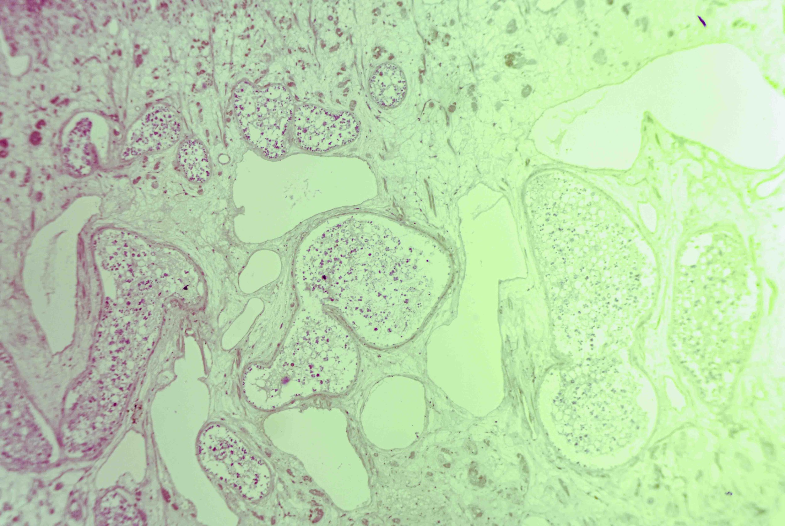
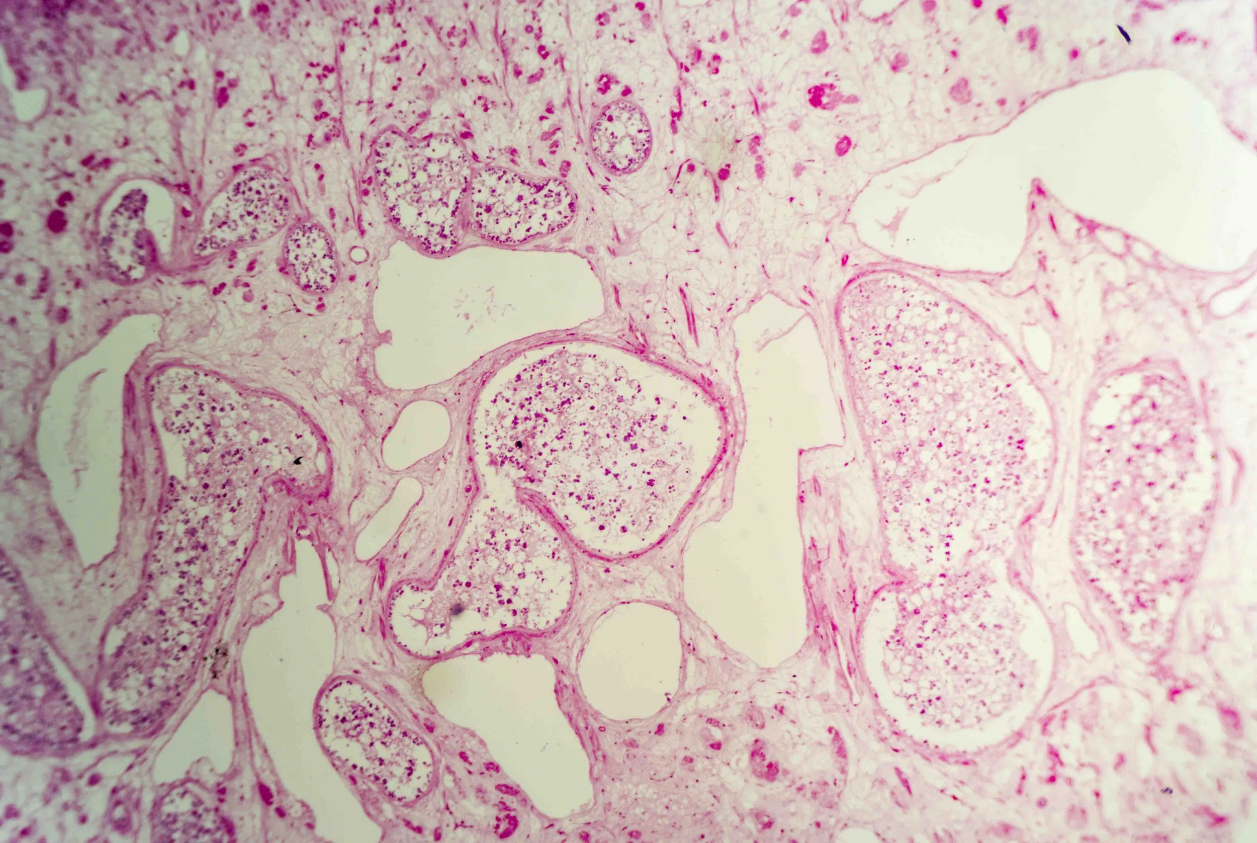
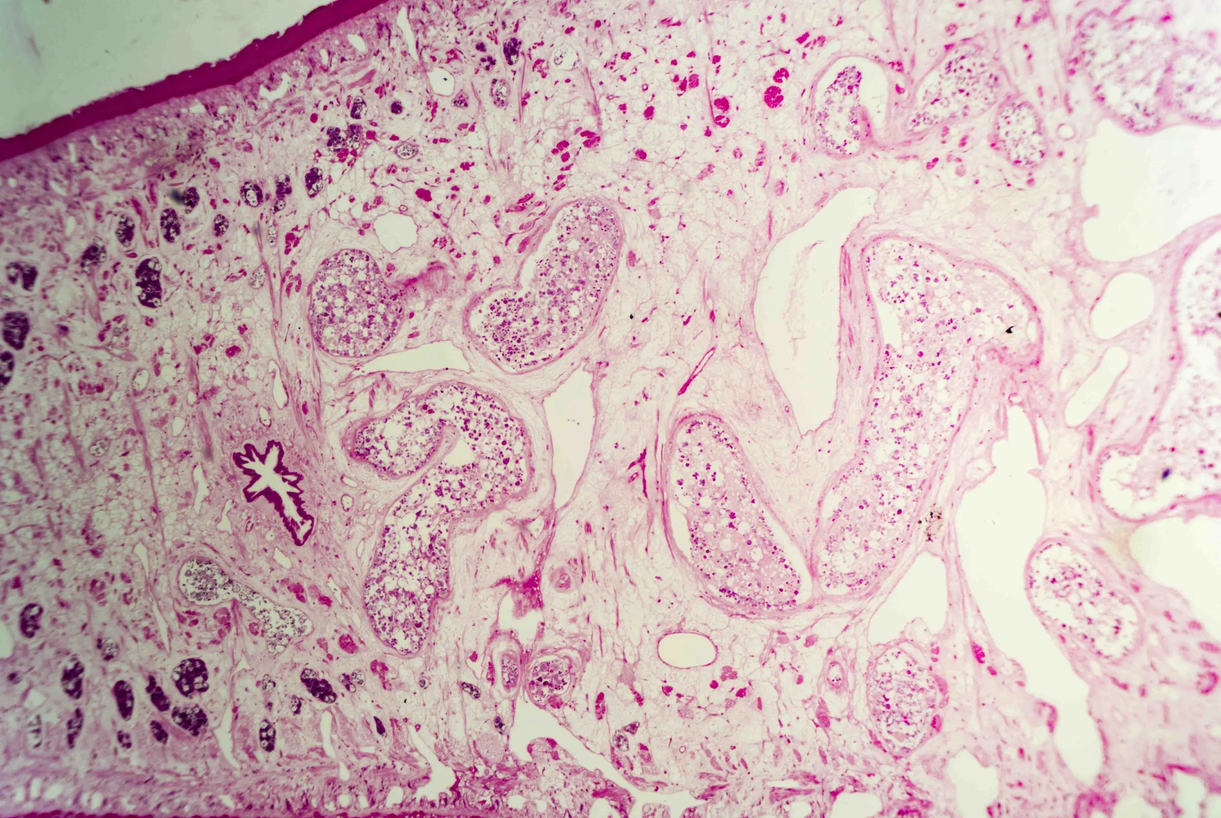
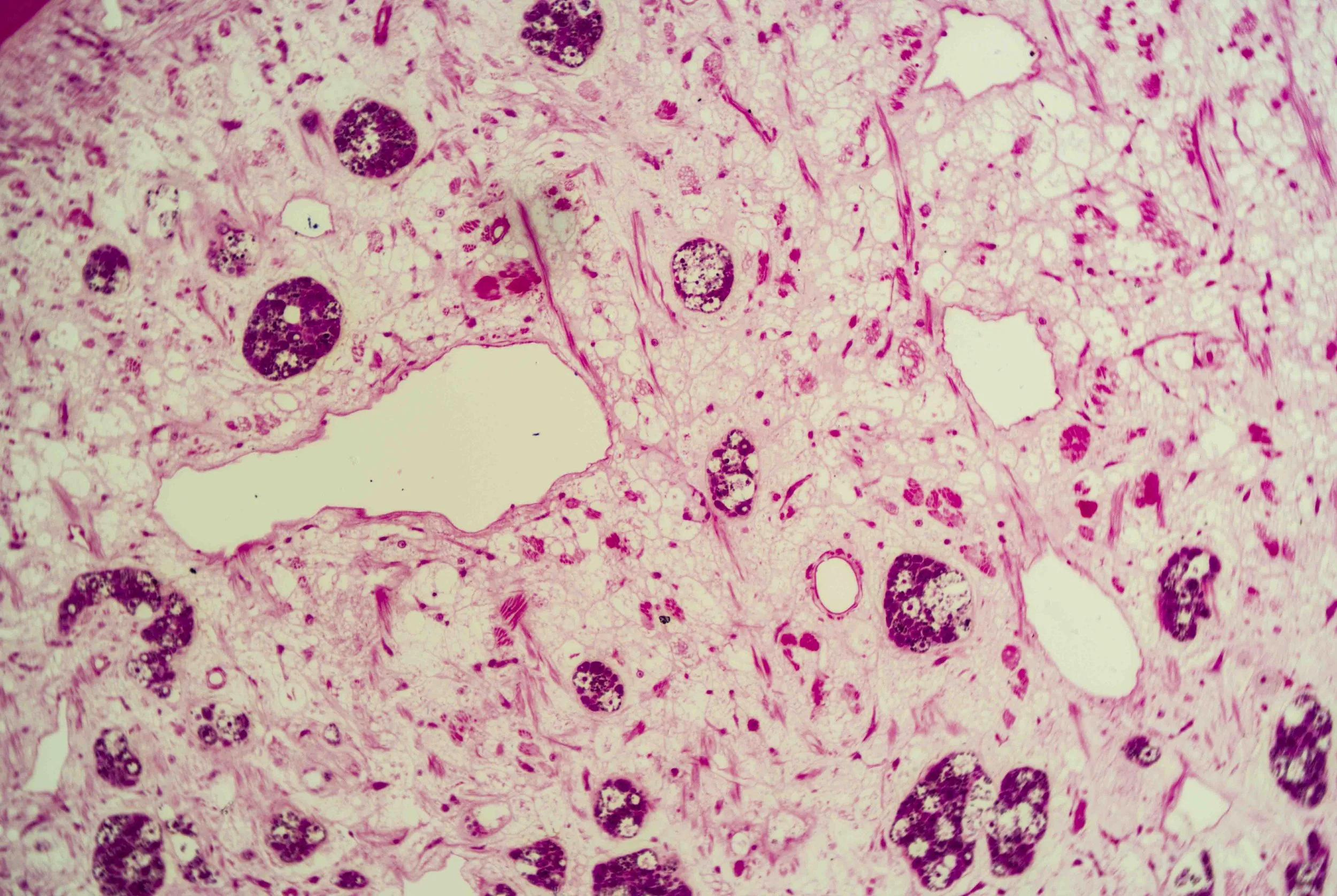
Hidden architecture of a parasite

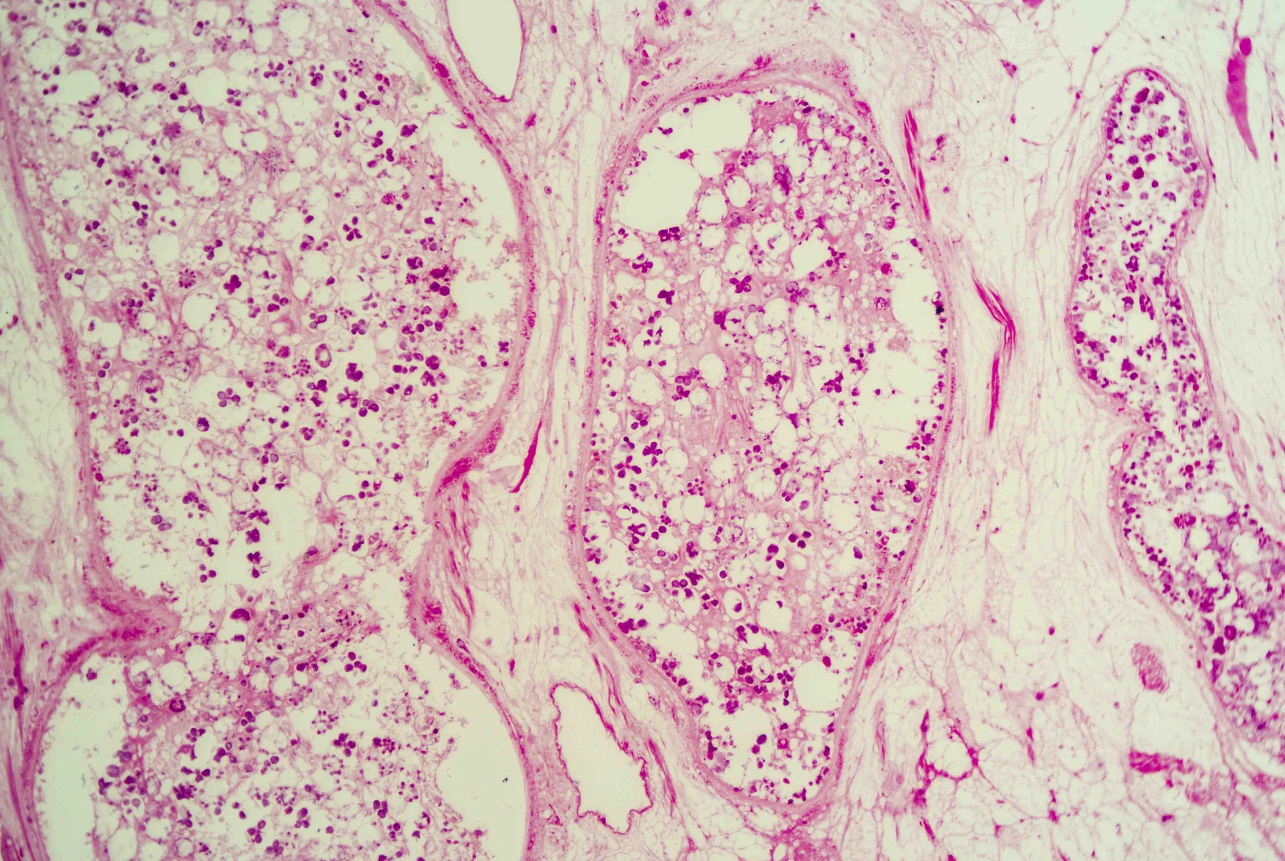
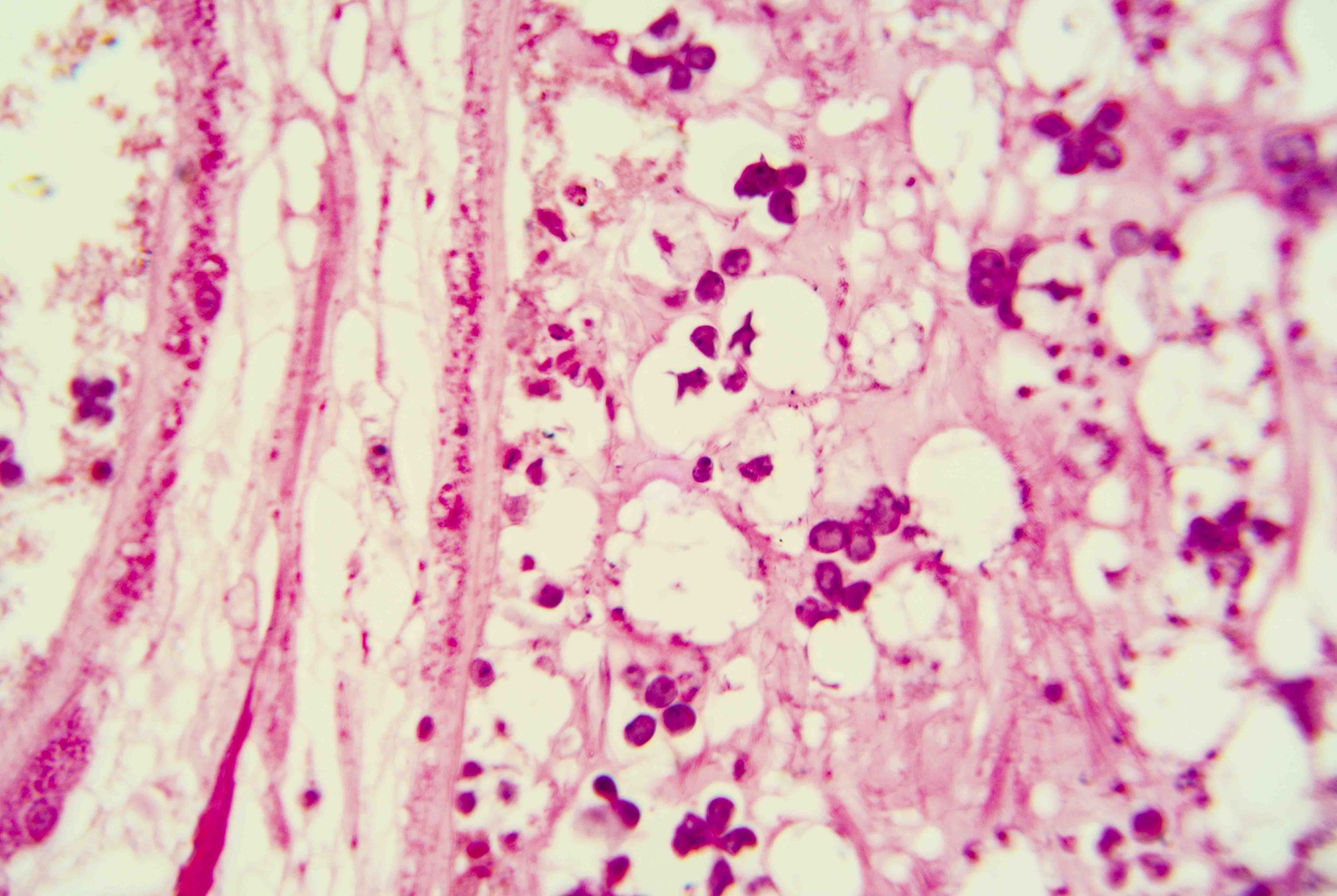
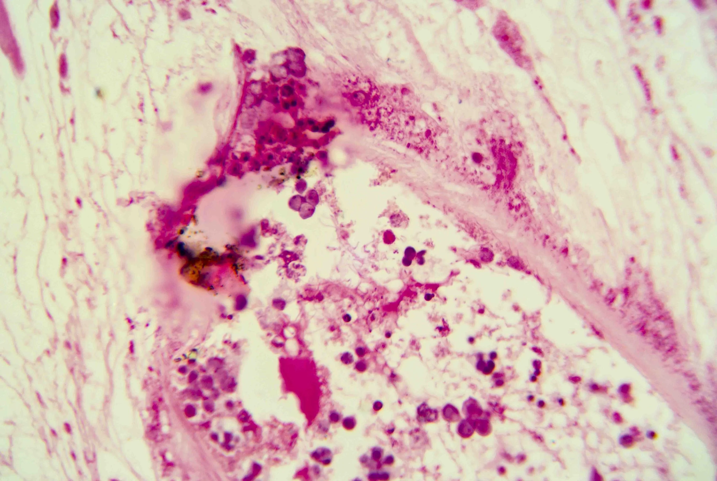
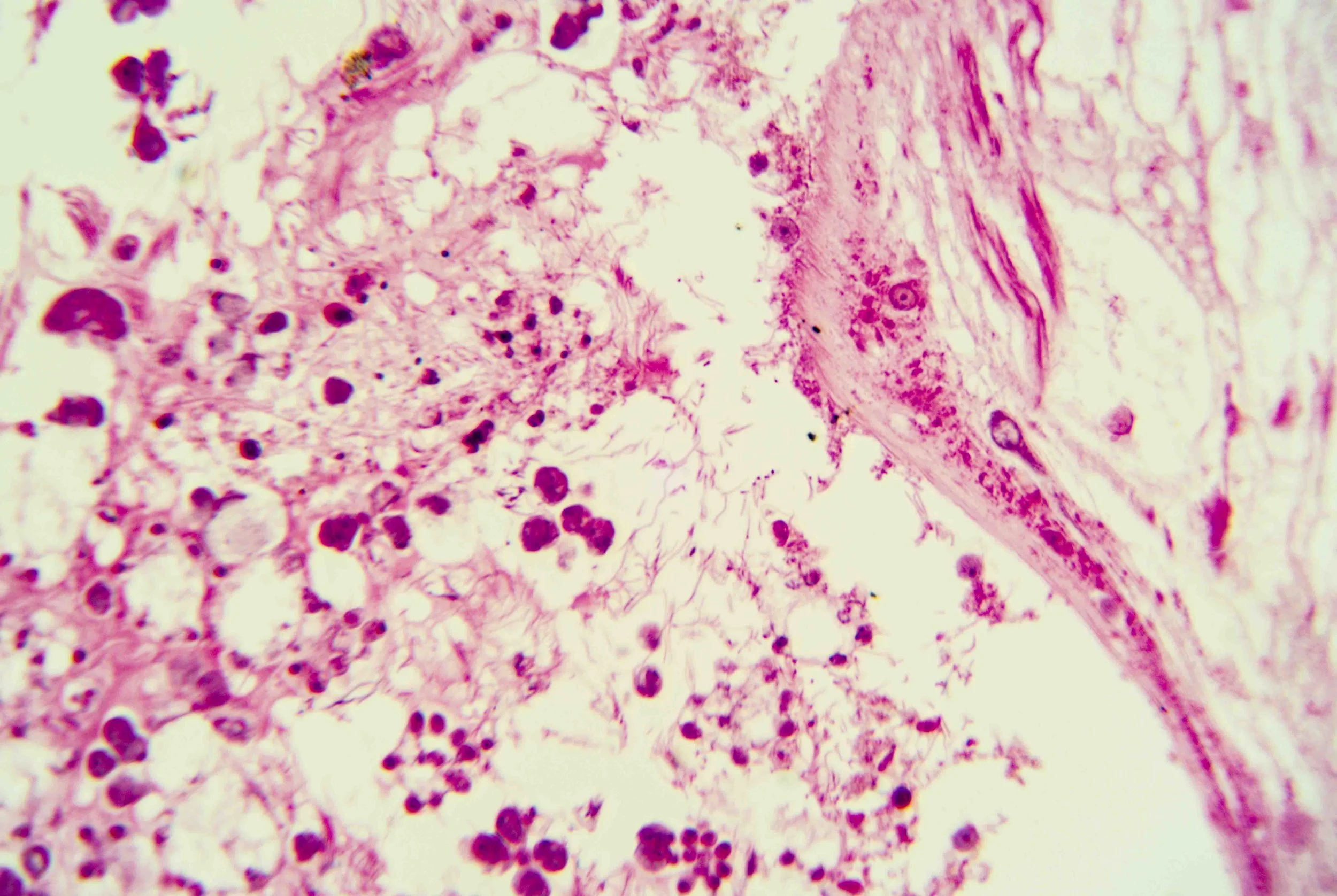
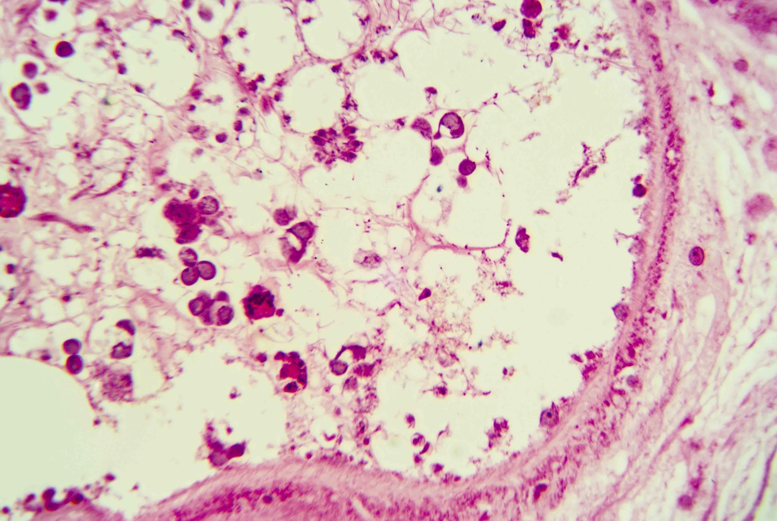
At first glance, a liver fluke is a simple, leaf‑shaped flatworm. Under magnification, however, a cross‑section reveals a labyrinth of chambers and cells, like stained‑glass windows filled with pink and purple mosaics. These sections show the parasite’s densely packed tissues, reproductive organs and digestive sacs nestled within its protective tegument.

This creature’s body is dorsoventrally flattened and oval, resembling a faint brown leaf. Adult flukes can reach about 3 centimetres in length and 1.5 centimetres in width, with a broader front tapering to a blunt posterior.

The anterior end forms a conical “head” bearing a small yet powerful oral sucker used for feeding and adhesion. Just behind it lies a larger ventral sucker (acetabulum) that anchors the fluke to its host’s tissues.

Unlike many animals, liver flukes lack an anus; their branched gut empties through the mouth.

The surface of the fluke is wrapped in a syncytial tegument—a scleroprotein‑rich skin enveloped in glycocalyx—that protects the parasite from digestive enzymes and allows nutrient absorption.

Tiny spines on this tegument become more numerous and multi‑pointed as the fluke approaches the bile ducts, helping it anchor and resist the flow of bile.

Liver flukes are hermaphrodites, carrying both male and female reproductive organs. They feed primarily on blood within the host’s liver and bile ductsen.wikipedia.org, and their branched intestine distributes nutrients throughout the body.

Eggs produced within the fluke exit through an excretory pore and hatch into larvae that infect freshwater snails. In many species, the life cycle requires multiple hosts: larvae develop in snails, sometimes encyst in fish, and finally mature in the liver or bile ducts of mammals—including humans—when contaminated vegetation or fish are consumeden.wikipedia.org.

These parasites cause fasciolosis in livestock and humans, a disease that can lead to fever, abdominal pain, jaundice and anaemia. Chronic infections with certain species, such as Opisthorchis viverrini and Clonorchis sinensis, are linked to long‑term inflammation and increased risk of bile‑duct cancer.

Yet there is an eerie beauty in their design. The intricate patterns seen in histological sections hint at the fluke’s complex adaptations—suckers, tegument, branched gut and prolific reproductive tissues—all evolved to exploit its ecological niche.

Observing these structures up close invites us to appreciate both the biological ingenuity of parasites and the hidden architectures that influence human and animal health.

Previous
Previous

Human Saliva

Next
Next

Mosquitoes