
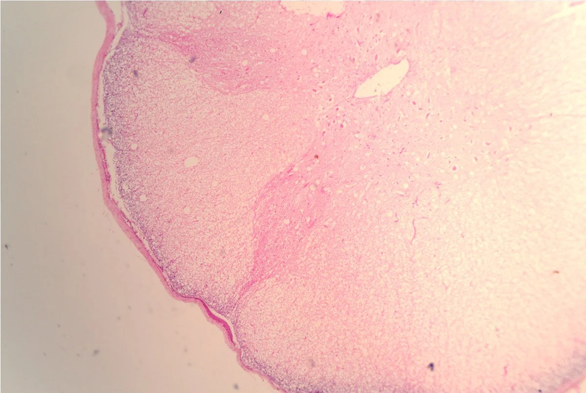
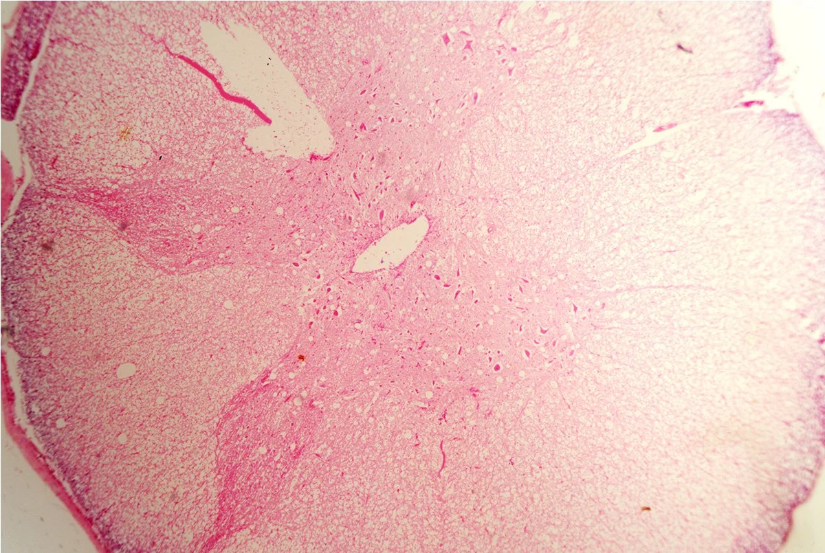
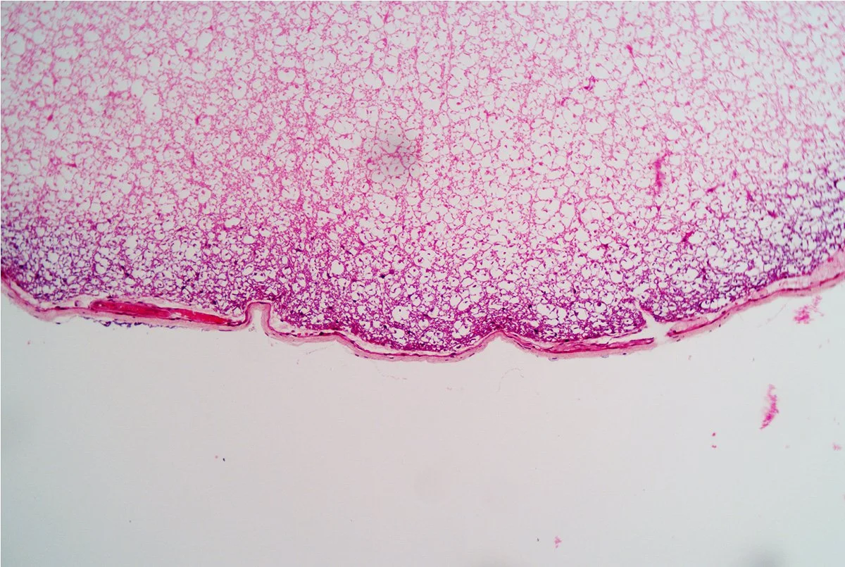
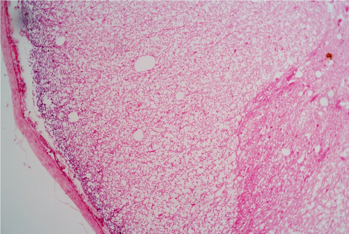
Seen through the microscope, the spinal cord reveals an elegant and highly organized structure.
In cross-section, its familiar butterfly-shaped gray matter sits at the center, surrounded by white matter tracts that carry electrical signals between the brain and the rest of the body.
These pathways coordinate movement, transmit sensation, and regulate countless automatic functions that sustain life.
Spinal Cord CS


The scientific understanding of the spinal cord developed gradually during the nineteenth century, when advances in microscopy allowed anatomists to explore the nervous system with increasing precision.
One of the most influential figures in this field was Santiago Ramón y Cajal, the Spanish neuroscientist who used refined staining techniques to visualize individual neurons. His work helped establish the neuron doctrine, demonstrating that the nervous system is composed of distinct cells that communicate through specialized connections. Cajal’s detailed drawings of neural tissue transformed neuroscience and earned him the 1906 Nobel Prize in Physiology or Medicine.
As scientists began to understand the structure of the spinal cord, they also recognized its vulnerability to infectious disease. In the early twentieth century, poliomyelitis became one of the most feared neurological illnesses in the world. The poliovirus selectively targeted motor neurons in the spinal cord, sometimes leaving survivors with lifelong paralysis. Major epidemics swept across Europe and North America during the first half of the century, driving an urgent global effort to understand the virus and protect the nervous system.
A turning point came in the 1950s when Jonas Salk developed the first widely used polio vaccine, followed later by the oral vaccine pioneered by Albert Sabin. Mass vaccination campaigns dramatically reduced the incidence of polio worldwide, transforming a once-common neurological catastrophe into a largely preventable disease.
Today, the spinal cord remains a central focus of medical research. Scientists continue to investigate how infections, inflammation, and autoimmune conditions affect this delicate network of neurons and glial cells. At the same time, advances in neuroimaging, molecular diagnostics, and regenerative medicine are offering new possibilities for protecting and repairing spinal tissue.
Under magnification, the spinal cord’s internal architecture appears almost artistic—curving tracts, branching neurons, and layered tissues arranged with remarkable precision. Yet this intricate structure is also a testament to the progress of neuroscience and medicine. From early microscopic observations to modern vaccines and neurological research, our understanding of the spinal cord reflects humanity’s ongoing effort to preserve one of the body’s most vital communication pathways.
In this gallery, the spinal cord becomes more than an anatomical structure. It is a reminder that scientific curiosity, careful observation, and collaborative discovery continue to illuminate the pathways that connect the brain, the body, and the microscopic world around us.